Radiologist
Dr. Nishant Mishra
Radiologist | Assistant Professor | Medical Educator
MBBS – IMS, Banaras Hindu University
MD Radiodiagnosis – PGIMS, Rohtak
Former Senior Resident – AIIMS Rishikesh & Lady Hardinge Medical College, New Delhi
Current: Assistant Professor, Department of Radiodiagnosis, NC Medical College
About Me
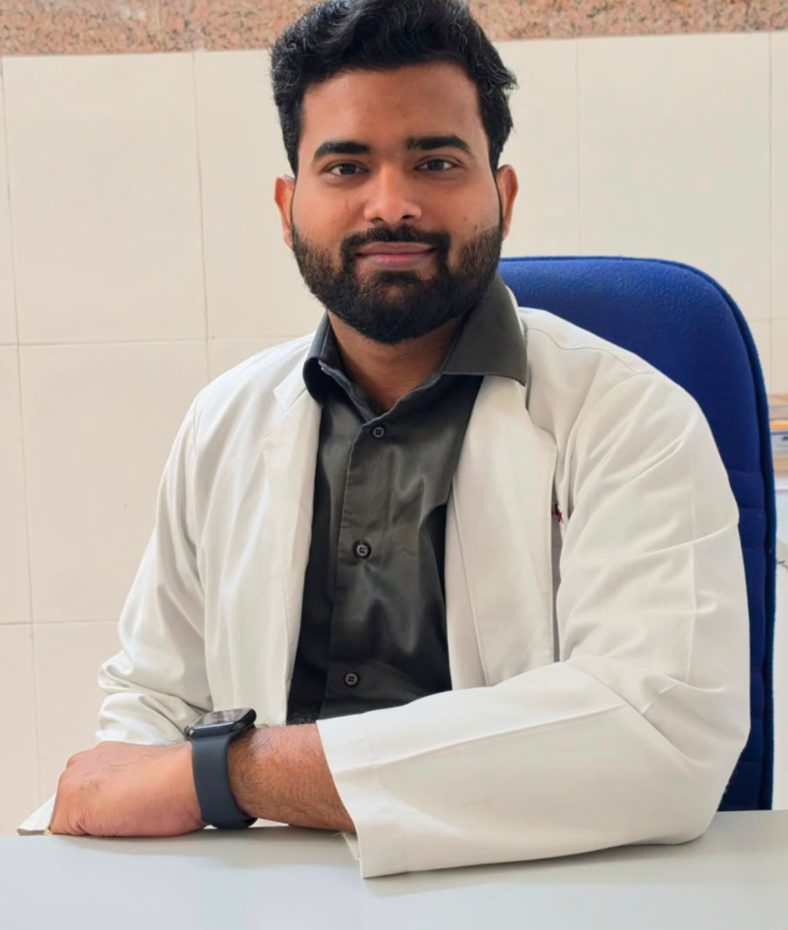
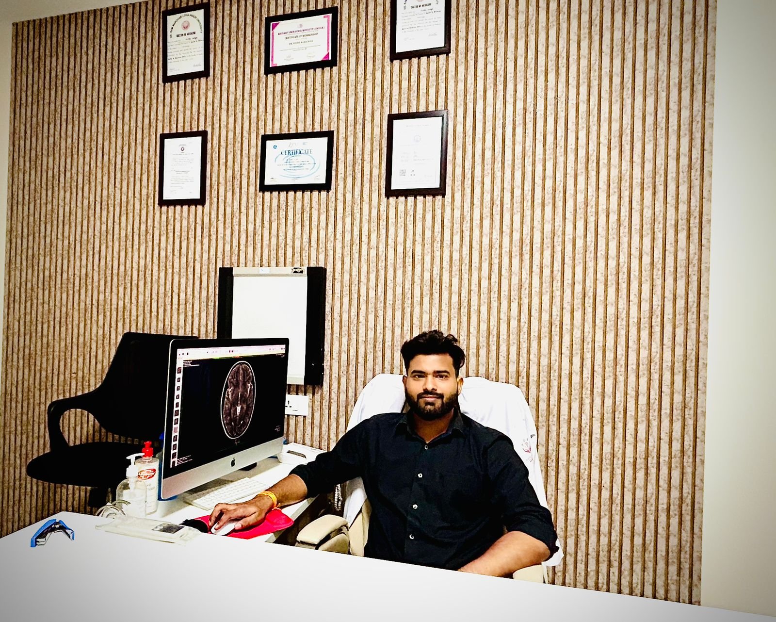
Dr. Nishant Mishra
Dr. Nishant Mishra is a Radiologist with 7+ years of expertise and dedication. He has extensive experience in the field and is committed to providing the best possible care to his patients. Dr. Mishra stays up-to-date with the latest advancements in radiology through regular attendance at CMEs, workshops, and national conferences, where he actively participates in presenting papers and posters.
He is a member of several prestigious medical organizations, including the Indian Radiological Imaging Society, Telemedicine Society of India, and National Medicos Organisation. In his spare time, Dr. Mishra enjoys traveling, playing chess, and volunteering to help those in need.
Academic Qualifications
- MBBS from the Institute of Medical Sciences, Banaras Hindu University in December 2018 (passed all professional exams on the first attempt)
- Internship training at Sundar Lal Hospital, Banaras Hindu University, UP(January 2019 to December 2020)
- Served as Telemedicine Medical Officer in the Regional Resource Centre (RRC), IMS, BHU under Gov. Of India from 8 February to 8 July 2020 (During Covid 1st wave)
- MD Radiodiagnosis from Pt. B. D Sharma, PGIMS Rohtak(completion date: July 12th, 2023)
- Senior Resident Doctor, KM, MCH, Mathura (13 July 2023 till date)
Professional Experience
NC Medical College
(Current)
Post : Assistant Professor

Lady Hardinge Medical College, New Delhi
Post : Senior Resident
AIIMS,
Rishikesh
Post : Senior Resident
K.M. Medical College, Mathura
Post : Senior Resident
One-Stop Solution for Radiology and General Practice
Dr. Nishant Mishra is a board-certified radiologist who also offers routine checkups, preventive care, chronic disease management, and treatment for acute illnesses and injuries.
Our Services
My Expertise
As a highly trained medical specialist, Dr. Nishant Mishra possesses extensive medical knowledge and advanced analytical skills in interpreting a wide range of diagnostic imaging tests.
Fetal Imaging
Conventional radiology is the use of X-rays to create images of the body’s internal structures. It allows professionals to diagnose and monitor various medical conditions.
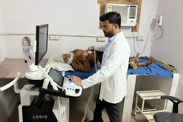
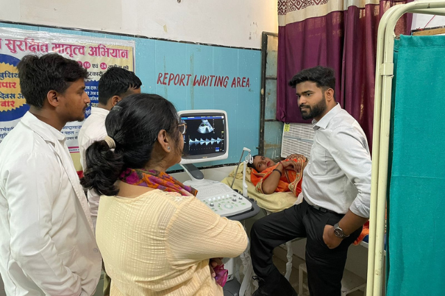
Obstetric Ultrasound
Ultrasonography is a medical imaging technique that uses high-frequency sound waves to create images of structures inside the body.
Gynecological Imaging
A Doppler study is a non-invasive test that uses sound waves to measure blood flow and detect any abnormalities in the heart or blood vessels.
Doppler Studies
A CT scan uses X-rays to create detailed images of the body’s internal structures, helping doctors diagnose and monitor various medical conditions.
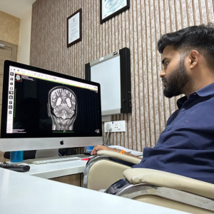
CT & MRI Interpretation
Interventional radiology uses imaging techniques to guide minimally invasive procedures, reducing recovery time and risks compared to traditional surgery.
Breast Imaging
MRI, or Magnetic Resonance Imaging, is a non-invasive medical procedure that uses powerful magnetic fields and radio waves to create detailed images of the body’s internal structures.
Ultrasound-Guided Procedures
A Doppler study is a non-invasive test that uses sound waves to measure blood flow and detect any abnormalities in the heart or blood vessels.
FAQs
Frequently Asked Questions

Preparing for a radiology exam depends on the type of test.
- Ultrasound: You might need to follow a special diet or take laxatives to clear your bowels. For pelvic ultrasounds, drink water beforehand to fill your bladder. Sometimes, fasting helps improve the images.
- Contrast Studies (CT or MRI): Fasting is usually required to avoid risks and get clearer images. Doctors will check your kidney function before using contrast agents.
Always tell your doctor about allergies and follow their instructions for the best results and safety.
Radiology exams like X-rays, CT scans, MRIs, and ultrasounds are generally safe when done by trained professionals. The main concern with X-rays and CT scans is the small risk of cancer from radiation, but doctors use low doses to keep patients safe. MRIs are very safe because they use magnets and radio waves instead of radiation. Ultrasounds use sound waves and are also safe, making them popular for many medical uses, especially in pregnancy. Overall, the benefits of these exams usually outweigh the risks, and doctors carefully choose the right test for each patient.
Radiological exams are designed to be as comfortable and easy as possible for patients. X-rays and CT scans involve lying still while the machine takes pictures. Ultrasound uses gel and sound waves to create images, without radiation or pain. MRI scans use magnets and radio waves, but earplugs reduce noise. Trained professionals perform these exams with patient comfort in mind. While each exam has its own process, discomfort is minimized through careful protocols and patient-centered care.
Lorem ipsum dolor sit amet, consectetur adipiscing elit. Integer nec odio. Praesent libero. Sed cursus ante dapibus diam. Sed nisi. Nulla quis sem at nibh elementum imperdiet. Duis sagittis ipsum. Praesent mauris.
Fusce nec tellus sed augue semper porta. Mauris massa. Vestibulum lacinia arcu eget nulla. Class aptent taciti sociosqu ad litora torquent per conubia nostra, per inceptos himenaeos. Curabitur sodales ligula in libero. Sed dignissim lacinia nunc.
Lorem ipsum dolor sit amet, consectetur adipiscing elit. Integer nec odio. Praesent libero. Sed cursus ante dapibus diam. Sed nisi. Nulla quis sem at nibh elementum imperdiet. Duis sagittis ipsum. Praesent mauris.
Fusce nec tellus sed augue semper porta. Mauris massa. Vestibulum lacinia arcu eget nulla. Class aptent taciti sociosqu ad litora torquent per conubia nostra, per inceptos himenaeos. Curabitur sodales ligula in libero. Sed dignissim lacinia nunc.