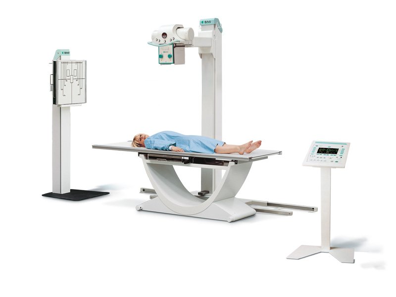
Conventional Radiology

Conventional radiology, also known as plain film radiography, is a type of medical imaging that uses X-rays to create images of the body’s internal structures. It is one of the oldest and most commonly used imaging techniques in healthcare.
In conventional radiology, a patient is positioned between an X-ray source and a film or digital detector. The X-rays pass through the body, and the resulting image is captured on the film or detector. Different tissues in the body absorb X-rays at different rates, creating a contrast between bones, soft tissues, and air spaces.
Conventional radiology is used to diagnose a wide range of conditions, including fractures, infections, and tumors. It is also used to guide certain medical procedures, such as the placement of catheters or needles.
Despite the development of more advanced imaging techniques, such as CT scans and MRI, conventional radiology remains an important tool in healthcare due to its speed, cost-effectiveness, and accessibility.
Different Types of Tests Included in Conventional Radiology
Barium Study
Barium study is a type of X-ray test that helps doctors see the shape and movement of your digestive tract. It uses a white, chalky liquid called barium to coat the inside of your esophagus, stomach, and intestines. This makes them show up clearly on the X-ray images. Doctors use barium studies to check for problems like blockages, tumors, or reflux.
IVP (Intravenous Pyelography)
IVP (Intravenous Pyelography) is a test that uses X-rays to take pictures of your kidneys, ureters, and bladder. It helps doctors see if there are any problems with these organs. During the test, a special dye is injected into your vein, which helps the X-rays show the organs more clearly. The test is usually done in a hospital or imaging center.
HSG / SSG
HSG (Hysterosalpingography) and SSG (Sonosalpingography) are imaging tests used to evaluate the fallopian tubes and uterus. HSG uses X-rays and a contrast dye, while SSG uses ultrasound and a saline solution. These tests can help diagnose blockages, abnormalities, or scarring in the fallopian tubes or uterus, which may cause infertility. The choice between HSG and SSG depends on individual factors and the preference of the doctor.
Fistueograu
Fistueograu, also known as fistulograms or fistulography, is a radiological procedure used to examine abnormal connections or passages called fistulas. During this test, a contrast dye is injected into the fistula, and X-rays are taken to see the path and extent of the fistula. This helps doctors diagnose and treat fistulas in various parts of the body.
T-Tube Cholangiography
T-Tube Cholangiography is a medical imaging technique used to examine the bile ducts after gallbladder surgery. A small tube called a T-tube is placed in the bile duct during the operation. Contrast dye is then injected through the T-tube, and X-rays are taken to create images of the bile ducts. This helps doctors check for any blockages or abnormalities.
Mammography
Mammography is a type of medical imaging that uses low-dose X-rays to examine the breasts. It helps detect breast cancer early, even before symptoms appear. The procedure involves placing the breast between two plates and taking X-ray pictures. Mammograms are capable of finding lumps or abnormalities that may be cancer. Regular mammograms are recommended for women over 40 to screen for breast cancer.
Digital Radiography
Digital radiography is a modern technique used in conventional radiology. It involves capturing X-ray images digitally, rather than on film. This allows for faster processing, enhanced image quality, and easier storage and sharing of images. Digital radiography is more efficient and eco-friendly compared to traditional film-based methods.
Fluoroscopy
Fluoroscopy is a type of medical imaging that uses X-rays to create real-time moving images of the body’s internal structures. It’s commonly used to guide procedures, such as inserting catheters or finding blockages in the digestive system. Fluoroscopy allows doctors to see what’s happening inside the body as they perform these tasks.
MCU /RGU
MCU (Micturating Cystourethrogram) and RGU (Retrograde Urethrogram) are X-ray imaging tests used to evaluate the urinary bladder and urethra. In an MCU, a small tube is inserted into the bladder to fill it with a contrast dye. X-rays after the test are then studied to check for any abnormalities. An RGU is similar, but the contrast dye is injected directly into the urethra.
Loopogram
A loopogram is a type of X-ray test that helps doctors see the small intestine. It uses a special dye that is injected into a small opening in the intestine. This dye helps doctors study the intestine clearly on the X-ray images. Doctors use loopograms to check for problems in the small intestine, such as blockages or tumors.