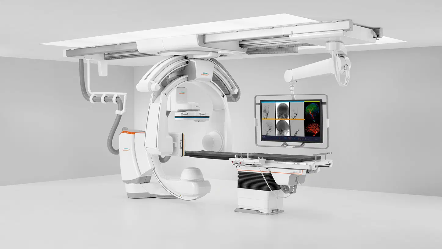
Interventional Radiology

Interventional Radiology (IR) is a medical specialty that uses imaging techniques like X-rays, CT scans, and ultrasound to guide minimally invasive procedures. IR doctors use small needles and catheters to treat various conditions without major surgery.
Some common IR procedures include:
- Embolization: Blocking blood flow to tumors or bleeding blood vessels
- Angioplasty: Opening blocked arteries with balloons and stents
- Biopsy: Taking tissue samples for diagnosis
- Drainage: Removing fluid from organs or abscesses
IR is often used to treat cancer, vascular diseases, and trauma. It can be a great alternative to many open surgery procedures. IR procedures are usually done with local anesthesia and sedation, so recovery time is faster than with traditional surgery.
IR is a rapidly evolving field that combines advanced imaging, minimally invasive techniques, and specialized expertise to provide effective, targeted treatments with less risk and faster recovery for patients.
Different Types of Tests Included in Interventional Radiology
Ultrasound Guided FNAC / Biopsy
Ultrasound Guided FNAC/Biopsy is a medical procedure done by radiologists. It uses sound waves to guide a thin needle to take a small sample of tissue or fluid from an area of concern. This helps diagnose conditions like cancer, infections, or other diseases. The procedure is minimally invasive and often done as an outpatient.
CT Guided Biopsy
A CT-guided biopsy is a minimally invasive procedure used to obtain a small sample of tissue from a specific area inside the body. It is performed using a CT scanner to guide a thin needle to the target site. The sample is then sent to a lab for analysis to diagnose conditions like cancer or infection.
USG - Guided Abscess Drainage / Pigtail Insertion
USG-Guided Abscess Drainage/Pigtail Insertion is a procedure done by radiologists to remove pus from an infected area. A small tube called a pigtail catheter is placed into the abscess using ultrasound guidance. The pus drains out through the tube, allowing the infection to heal. This minimally invasive procedure is often done instead of surgery.
USG - Guided Prostate Biopsy
Transrectal ultrasound-guided prostate biopsy is the standard approach for diagnosing prostate cancer. The doctor inserts a lubricated ultrasound probe into the rectum to guide a biopsy needle into the prostate to collect tissue samples. This procedure is usually done on an outpatient basis under local anesthesia. Minor complications like bleeding are common but serious infections are rare with proper antibiotic prophylaxis
USG - Guided Breast Biopsy
Ultrasound-guided breast biopsy is a minimally invasive procedure used to diagnose breast abnormalities. A radiologist uses ultrasound imaging to guide a needle into the suspicious area and collect tissue samples for examination under a microscope. This technique allows for precise targeting of the lesion and is often used for abnormalities that are not easily felt during a physical exam.
USG - Guided Kidney Biopsy
A kidney biopsy is a procedure where a small piece of tissue is removed from the kidney for testing. It’s done using ultrasound or CT guidance to ensure accuracy. The doctor numbs the area, then uses a special needle to take the sample. This helps diagnose kidney problems and guide treatment. It’s a safe procedure, but there may be some discomfort or bleeding afterwards.
Radio-Frequency Ablation of Varicose Vein
Radio-Frequency Ablation (RFA) is a procedure mainly used to treat varicose veins. It is a minimally invasive procedure that uses heat from radio waves to seal the damaged vein. A thin tube called a catheter is inserted into the vein through a small incision. Radio waves are then sent through the catheter to heat and close the vein. This allows blood to flow through healthier veins.
USG - Guided Ascitic & Pleural Tapping
USG-Guided Ascitic & Pleural Tapping is a procedure done by radiologists to remove fluid from the abdomen (ascites) or chest (pleural effusion). It uses ultrasound to guide a thin needle to the fluid area. This helps remove the fluid safely and accurately. The procedure is done to relieve symptoms and get fluid samples for testing.
Neuro Intervention
Neuro Intervention in Interventional Radiology refers to minimally invasive procedures that treat brain and nervous system conditions. These procedures use imaging guidance to access the problem area through small incisions. Neuro Interventional Radiology can treat strokes, aneurysms, tumors, and other issues without major surgery. It offers a less invasive option for many neurological conditions.
Peripheral Vascular Intervention
Peripheral Vascular Intervention (PVI) is a minimally invasive procedure performed by interventional radiologists to treat blocked or narrowed blood vessels in the legs, arms, and other parts of the body. PVI uses small catheters and imaging guidance to open up blocked arteries and improve blood flow. It is often used as an alternative to surgery for patients with peripheral artery disease (PAD).