MRI (Magnetic Resonance Imaging)

Magnetic Resonance Imaging (MRI) is a powerful tool used by doctors to see inside the human body without making any cuts. It uses strong magnets and radio waves to create detailed images of organs, tissues, and bones.
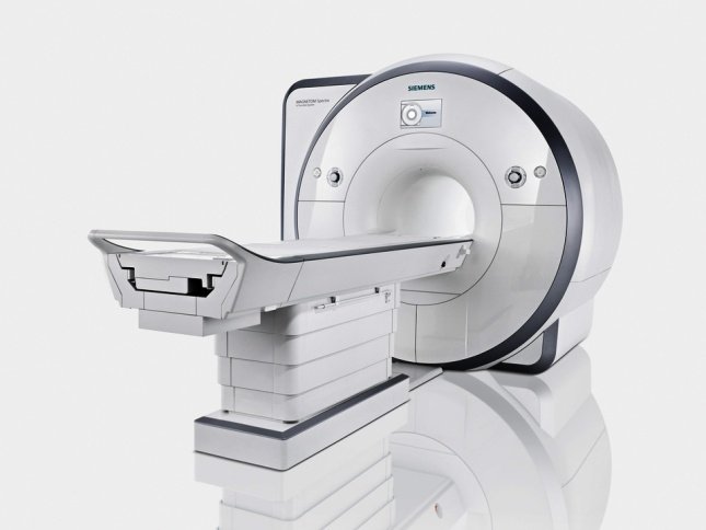
MRI machines are large, tube-shaped devices that patients lie inside during the scan. The machine takes pictures from different angles, allowing doctors to see problems like injuries, infections, or tumors. MRI scans are painless and safe, but they can be noisy and make some people feel claustrophobic.
Doctors use MRI scans to diagnose many health issues, including brain disorders, heart problems, and joint injuries. The images help them plan treatments and track how well patients are responding to therapy. MRI is an important part of modern healthcare, providing valuable information to keep people healthy.
Different Types of Tests Included in MRI (Magnetic Resonance Imaging)
ASL (Non Contrast Perfusion)
ASL (Arterial Spin Labeling) is a non-invasive MRI technique that measures blood flow in the brain without using contrast agents. It works by magnetically “labeling” the water protons in the blood, then detecting their signal as they flow into the brain. ASL provides information about brain function and can help diagnose conditions like stroke, tumors, and neurodegenerative diseases.
Functional MRI
Functional MRI (fMRI) is a type of MRI that measures brain activity. It uses magnetic fields and radio waves to detect any changes in the blood flow and oxygen levels in the brain. These changes are linked to specific brain functions, such as seeing, hearing, or thinking. fMRI helps doctors understand how the brain works and identify any abnormalities.
Tractography
Tractography is a technique used in magnetic resonance imaging (MRI) to visualize and map the brain’s white matter pathways. It uses diffusion-weighted imaging (DWI) to track the movement of water molecules within the brain, which can reveal the direction and organization of nerve fibers. This information helps doctors understand brain structure and function.
Cardiac MRI
Cardiac MRI is used to diagnose and identify heart conditions. It uses strong magnetic fields and radio waves to create detailed images of the heart. These images help doctors see how well the heart is working and identify any problems. Cardiac MRI is safe, painless, and does not use radiation like X-rays. It is an important part of heart health care.
Breast MRI
Breast MRI is a powerful tool for detecting and diagnosing breast cancer. It uses strong magnetic fields and radio waves to create detailed images of the breast tissue. Breast MRI can identify tumors that may be too small to be felt during a physical exam or seen on a mammogram. It is often used in addition to other breast imaging tests to get a more complete picture of a patient’s breast health.
Fetal MRI
Fetal MRI is a powerful tool that allows doctors to see detailed images of a baby inside the mother’s womb. It uses strong magnets and radio waves to create clear pictures of the baby’s body. Doctors can use these images to check for any problems or abnormalities. Fetal MRI is safe for both the mother and the baby.
Multiparametric Prostate MRI
Multiparametric prostate MRI is a powerful tool for detecting and diagnosing prostate cancer. It combines different MRI techniques to create detailed images of the prostate gland. This allows doctors to identify suspicious areas and determine the best course of treatment. Multiparametric MRI is becoming increasingly important in prostate cancer management, offering a more precise and less invasive approach compared to traditional methods.
Liver MRI
Liver MRI is a medical imaging technique that uses strong magnetic fields and radio waves to create detailed images of the liver. It helps doctors see if there are any abnormalities or problems in the liver. The MRI machine is a large, tube-shaped machine that the patient lies inside during the scan. Liver MRI is a safe and painless procedure that can provide important information about the liver’s health.
MR Enterography
MR Enterography is a type of magnetic resonance imaging (MRI) that allows doctors to see detailed images of the small intestine. Patients are instructed to drink a special contrast liquid before the scan. This liquid helps make the intestines show up clearly on the MRI images. MR Enterography can help diagnose problems like Crohn’s disease, tumors, or blockages in the small intestine.
MR Neurography
MR Neurography is a specialized MRI technique that provides detailed images of nerves in the body. It helps doctors diagnose nerve-related conditions like injuries, tumors, or compression. MR Neurography uses strong magnetic fields and radio waves to create clear pictures of nerves, allowing doctors to see if there are any problems or abnormalities
Defecography
Defecography in MRI is a technique that helps doctors understand how your bowels work. It uses special MRI scans to take pictures of your body while you have a bowel movement. This helps doctors see if there are any problems with how your bowels move or if there are any blockages. The test is usually done if you have trouble having bowel movements or have pain when you go to the bathroom.
Comprehensive Epilepsy Judging
Comprehensive Epilepsy Judging in Magnetic Resonance Imaging (MRI) is a technique used to analyze brain scans of people with epilepsy. It helps doctors identify abnormalities in the brain that may be causing seizures. This method uses advanced MRI technology to create detailed images of the brain, which are then carefully examined by experts to diagnose and treat epilepsy effectively.
MR Spectroscopy
MR Spectroscopy is a powerful technique used in Magnetic Resonance Imaging (MRI) to analyze the chemical composition of tissues. It measures the concentration of specific molecules in the body, helping doctors diagnose and monitor various medical conditions, such as brain tumors, Alzheimer’s disease, and heart disease. By understanding the chemical makeup of tissues, MR Spectroscopy provides valuable insights into the body’s health.
MR Arthrogram
An MR Arthrogram is a type of MRI scan that uses a contrast agent to get a clearer picture of the joints. The contrast agent is injected into the joint, and then the MRI machine takes images. This helps doctors see if there are any problems with the joint, like tears or other damage.
Spine DNI
Spine DNI in MRI stands for Spine Degenerative and Non-Inflammatory conditions. It refers to the use of magnetic resonance imaging (MRI) to detect and diagnose various spine problems that are not caused by inflammation or infection. MRI helps doctors see the spine’s structure and identify issues like disc herniation, spinal stenosis, and degenerative disc disease.
Brachial and Lumbosacral Plexus Imaging
Brachial and lumbosacral plexus imaging in MRI helps doctors see the nerves in your arms and legs. It uses powerful magnets and radio waves to create detailed pictures of these nerves. This allows doctors to check for any problems or injuries. The test is completely painless and is usually done in about an hour.