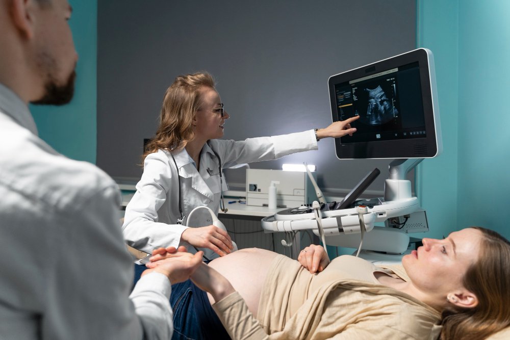
Ultrasonography

Ultrasonography, a non-invasive diagnostic procedure, employs high-frequency acoustic vibrations to generate visual representations of internal bodily components. It is a safe and non-invasive procedure that does not use radiation.
During an ultrasound, a small handheld device called a transducer is placed on the skin and moved around the area of interest. The transducer sends sound waves into the body, and the echoes that bounce back are used to create real-time images on a computer screen.
Ultrasonography is commonly used to examine the heart, blood vessels, kidneys, liver, and other organs. It is also used during pregnancy to monitor the development of the fetus and to check for any abnormalities.
The images produced by ultrasonography are used by doctors to diagnose and monitor various medical conditions, as well as to guide certain medical procedures, such as biopsies and injections.
Different Types of Tests Included in Ultrasonography
USG-Chest, Neck
Chest and neck ultrasound (USG) is a valuable tool for imaging these areas. It uses high-frequency sound waves to create images of the lungs, heart, and surrounding structures. USG can detect fluid buildup, tumors, and other abnormalities. It is often used to guide biopsies and fluid removal procedures. Compared to X-rays and CT scans, USG is radiation-free, portable, and can be done at the bedside.
MSK USG
Musculoskeletal ultrasound (MSK USG) is a non-invasive imaging technique that uses sound waves to create pictures of muscles, tendons, ligaments, and joints. It helps doctors diagnose and treat conditions like muscle tears, tendinitis, arthritis, and nerve problems. MSK USG is safe, quick, and doesn’t use radiation. A handheld probe is moved over the skin to obtain the images.
Sonomommography
Sonomammography is a type of ultrasound imaging used to examine the breast. It uses sound waves to create pictures of the breast tissue. This procedure can help detect lumps or abnormalities that may not be visible on a mammogram. Sonomammography is often used as a follow-up test after an abnormal mammogram result.
Level-I (NT / NB Scan)
Level-I (NT/NB Scan) in ultrasonography is a simple test used to measure the thickness of the skin at the back of a baby’s neck. This measurement helps detect potential chromosomal abnormalities, such as Down syndrome, during pregnancy. The test is usually performed between weeks 11 and 13 of pregnancy and takes only a few minutes.
II (Anomaly Scan)
II (Anomaly Scan) is a specialized ultrasound examination that looks for any abnormalities or problems in the fetus during pregnancy. It is typically performed between 18-22 weeks of gestation. The scan checks the baby’s organs, limbs, and overall development. It helps identify potential issues early on, allowing for proper monitoring and care.
Follicular Monitoring
Follicular monitoring is a technique used in fertility treatments to track the development of follicles in a woman’s ovaries. Follicles are fluid-filled sacs that contain eggs. Ultrasound scans are used to measure the size and number of follicles. This information helps doctors determine the best time for egg retrieval or insemination. Regular monitoring ensures the treatment is progressing as planned.
Fetal Echo
Fetal Echo is a type of ultrasound that examines the baby’s heart during pregnancy. It helps doctors check for any heart problems or abnormalities. The test is usually done between weeks 18 and 22 of pregnancy. It’s a safe and painless procedure that uses sound waves to create images of the baby’s heart.
USG - OBS Routine Monitoring
USG – OBS Routine Monitoring is a technique used during pregnancy to check the baby’s growth and development. It uses sound waves to create images of the baby in the mother’s womb. This helps doctors monitor the baby’s health and detect any potential issues early on.
USG - Abdomen & Pelvis
Ultrasound imaging of the abdomen and pelvis is a non-invasive way to see organs and structures inside your body. It uses sound waves to create pictures. The test can help diagnose problems like gallstones, kidney stones, tumors, or infections. It’s often used to guide procedures like biopsies or to check on a baby during pregnancy.
USG - Pelvis / RPOC
USG – Pelvis / RPOC is an ultrasound exam that checks for retained placental tissue after childbirth. It helps doctors see if any placenta is left in the uterus. This is important because leftover tissue can cause bleeding or infection. The exam is quick and painless. It uses sound waves to create images of the uterus on a screen.
KUB Scan (Kidney Ureter Bladder Scan)
A KUB scan is an ultrasound examination of the kidneys, ureters, and bladder. It uses sound waves to create images of these organs, helping doctors check for any abnormalities or problems. The procedure is quick, painless, and safe. It doesn’t use radiation, making it suitable for pregnant women and children.
USG - Cranium
Cranial ultrasound (CUS) is a valuable tool for evaluating a baby’s brain during the first year of life. It uses sound waves to create images of the brain, cerebrospinal fluid, and surrounding structures. CUS is portable, inexpensive, and can be done frequently without radiation exposure. It is often used to check for bleeding, brain damage, or abnormal development in premature or sick infants.
USG - Pelvis / RPOC
USG – Pelvis / RPOC is an ultrasound exam that checks for retained placental tissue after childbirth. It helps doctors see if any placenta is left in the uterus. This is important because leftover tissue can cause bleeding or infection. The exam is quick and painless. It uses sound waves to create images of the uterus on a screen.
KUB Scan (Kidney Ureter Bladder Scan)
A KUB scan is an ultrasound examination of the kidneys, ureters, and bladder. It uses sound waves to create images of these organs, helping doctors check for any abnormalities or problems. The procedure is quick, painless, and safe. It doesn’t use radiation, making it suitable for pregnant women and children.